
JERUSALEM — Researchers have developed an innovative AI-powered method to drastically improve the speed and accuracy of cancer Magnetic Resonance Imaging (MRI) scans, transforming how clinicians track tumor development in real time.
The breakthrough technology, named ELITE, was detailed in the journal Nature Communications and announced in a statement by the Israel Institute of Technology. While specifically engineered and tested for breast cancer imaging, the developers noted that the methodology holds the potential to be applied to brain, head, and neck scans, alongside other medical imaging platforms in the future.
Overcoming the Temporal Bottleneck of Traditional MRIs
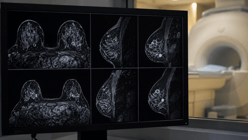
Dynamic MRI exams are highly valued for their detailed tissue visualization, making them a clinical standard for detecting breast cancer in high-risk populations. However, traditional systems are severely constrained by time, typically generating only one image every one to two minutes. This slow capture rate creates a significant blind spot, making it exceptionally difficult for radiologists to track how contrast materials move through biological tissue in real time.
The research team bypassed this technological bottleneck by fusing advanced mathematical modeling with a deep-learning system. This specialized AI pipeline is trained to simultaneously eliminate image noise and reconstruct missing spatial data.
The integration allows the system to produce high-quality scans at a staggering rate of one image per second, entirely eliminating the data lag associated with conventional scanning methods.
Enhanced Visibility in Clinical Trials
In clinical experiments evaluating 54 patients, the ELITE technology demonstrated a substantial leap in diagnostic performance, showing vastly improved tumor visibility and higher diagnostic sensitivity when compared directly with existing imaging protocols.
Medical experts conclude that the capability to track contrast agents almost continuously through tissue will allow doctors to identify micro-tumors much earlier. Crucially, this real-time tracking behavior provides the highly detailed kinetic data required to more accurately differentiate between benign and malignant growths, potentially reducing unnecessary biopsies and accelerating targeted treatment paths.